Tumour Imaging
The purpose of imaging is to directly observe where something goes, how it gets there, and how long it stays there. This holds true for anything from small molecules, viral vectors, to even the elusive cancer stem cells. There are two ways to image; invasively and non-invasively. The advantages of non-invasive imaging include rapid image acquisition, instantaneous information on biodistribution and kinetics, and most importantly, not having to sacrifice an animal for each time point. However this does not come without a price, and that price to be paid is level of detail. Invasive imaging involves removal of tissues of interest for imaging under either wide field or high magnification microscopy. Invasive imaging permits a level of detail down to the sub-cellular level, which captures the microenvironment of the tumour that is otherwise overlooked.
In the Liu lab, we perform both types of imaging to track the biodistribution and kinetics of the therapeutic compounds that we use.
For adenoviral work, we utilize bioluminescence imaging, which involves the collection of luciferase-mediated chemiluminescent photons by what is essentially a very sensitive digital camera.
For vesicular stomatitis virus (VSV) work, we utilize fluorescence imaging, which involves the excitation of fluorescent proteins produced by VSV with a bright blue light, which in turn emits green light, that is recorded by a wide-field stereomicroscope.
When investigating small molecules such as antisense oligonucleotides, we first conjugate them to a fluorescent molecule, such as FITC, then examine its microdistribution patterns following the excision of tissues of interest.
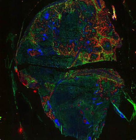
FITC-conjugated
antisense distribution in a C15 tumour
Red: active blood vessels, Green: antisense, Blue: hypoxia
Representative Publications:
|